Peripheral Venous Thrombolysis (Clot-Busting)
The term venous thromboembolism (VTE) is used to describe two conditions, deep vein thrombosis (DVT) and pulmonary embolism (PE). The term is used because the two conditions are very closely related. And because their prevention and treatment are also closely related.
-
Peripheral deep vein thrombosis. This is a blood clot that forms in a vein in the leg or arm. It can block blood flow. This can cause swelling, redness, and pain. These clots need treatment right away to prevent serious complications, like a blood clot in the lungs.
-
Pulmonary embolism. A piece of the clot may break off and travel to the lungs (pulmonary embolism). This is very serious and may cause death. The symptoms are chest pain, trouble breathing, coughing (may cough up blood), a fast heartbeat, sweating, and fainting. If you have these symptoms, call 911 or get emergency help.
Peripheral venous thrombolysis is a procedure to dissolve a blood clot in a leg or arm vein. This lessens the pain, redness, swelling, and other symptoms and prevents pulmonary embolism. The procedure is often done by a specialist called an interventional radiologist.
Before the procedure
Follow any instructions you are given on how to get ready. These include:
-
Directions you’re given for not eating or drinking before the procedure.
-
Making sure the healthcare providers know all of the medicines you take. This includes over-the-counter medicines, herbs, and supplements. The healthcare providers must also know if you are or may be pregnant. And if you are allergic to X-ray dye (contrast medium) or other medicines.
During the procedure
-
You will be carefully watched during the procedure.
-
You will be given fluids and medicines through your vein (intravenous or IV). You will be given medicine to help you relax and make you sleepy (sedation). You will also receive numbing medicine (local anesthesia).
-
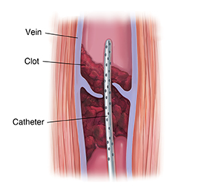
A very small cut (incision) is made in the area near the clot, like behind the knee. A thin, flexible tube (catheter) is put through the incision into the vein.
-
Contrast medium is injected through the catheter into the vein. This helps the vein show clearly on X-ray images. The radiologist uses these images as a guide. The radiologist moves the catheter through the vein to the clot.
-
When the catheter reaches the clot, the radiologist adds medicine or uses a device to dissolve the clot. If medicine is used, this is done slowly, over a period of a few hours. The catheter is left in place until the clot has dissolved. This can take up to 72 hours.
-
When the procedure is finished, the catheter is taken out. A healthcare provider will apply pressure to stop bleeding.
After the procedure
-
You will stay still for a period of time.
-
You will probably stay in the hospital for a few days and will continue to be carefully watched.
-
Drink a lot of water and other healthy drinks to help flush the contrast medium from your system.
-
After you go home, make sure you follow all instructions for taking care of your incision.
Possible risks and complications
-
Bleeding within the body or brain, or at the site where the catheter was placed
-
Damage to the vein or nearby structures
-
Infection
-
Problems because of contrast medium, including allergic reaction or kidney damage
© 2000-2024 The StayWell Company, LLC. All rights reserved. This information is not intended as a substitute for professional medical care. Always follow your healthcare professional's instructions.